The History of SpermatoStages™
⬇ Download PDFUnderstanding Spermatogenesis
Necessity is the mother of invention, and that proved true in the development of software to model the stages of spermatogenesis. In 1983, I began postdoctoral training at the U.S. Environmental Protection Agency (EPA; Research Triangle Park, NC) in the Reproductive Toxicology Branch of the Developmental Toxicology Division. I was assigned the task of understanding the stages of spermatogenesis, because most of the Branch’s focus had been developmental biology and female reproduction. At the time, the literature was sparse: PubMed lists only 53 papers on “spermatogenesis and stages” from 1950 to 1972, and just 293 papers from 1973 to 1983 (searched October 24, 2026). Original publications were hard to locate, buried in paper books and journals, and the published photographs were often of low quality. Matching what appeared under the microscope with the published descriptions was difficult, and the observed cellular transitions did not always align precisely with the literature. However, I was able to begin this long journey with encouragement from outstanding scientists at the EPA (including L. Earl Gray, John Laskey, and Ralph Linder) and a visit with R.E. (Bob) Chapin at the Chemical Industry Institute of Toxicology (CIIT), who was developing high resolution morphology methods for stage identification. Bob was one of the first to recognize the importance of understanding stages for pathological assessment of testicular effects in toxicology (Chapin et al. 1983, Chapin et al. 1984). He kindly taught me how to identify stages in the rat testis.
The Question
In July of 1986, I accepted an Assistant Professorship in the College of Veterinary Medicine at the University of Illinois at Urbana-Champaign. My responsibilities included teaching histology and doing research in male reproductive biology and toxicology. Fortunately, I was able to continue some of the EPA research that I was collaborating on. During the next two years, between lectures and setting up a lab, I spent many hours perfecting histology methods and looking through the microscope, trying to make sense of the staging skills passed on by Bob Chapin. Unfortunately, what I was seeing in testicular cross-sections was not always so precise; cells were frequently in transition from one stage to the next, making staging more difficult than often presented in the literature. I began to tabulate these differences and eventually decided to publish the data, with the hope of helping others with stage identification problems (Hess 1990). Toxicology also added to this difficulty, as pathological changes in testes often made staging decisions uncertain. However, an even more important question was raised, even while I was at the EPA: “What was the target cell type affected by the initial chemical treatment, if at a specific time post-exposure we observe pathological effects on a specific germ cell?”
Collection of Data
To answer ‘The Question’ required more than stage identification. The answer lay in another type of data, the kinetics of spermatogenesis or the ability to trace germ-cell development across all the stages in time and in the different target species. Surprisingly, the kinetics of spermatogenesis (duration in time) had been published before 1980 for most research animals, some as early as the 1950s (Roosen-Runge 1952, Clermont et al. 1959). Therefore, I began another literature review, also laborious and difficult to comprehend. I could understand how they determined the duration of spermatogenesis, by injecting tritiated-thymidine and looking for silver grains in photographic emulsion-coated histology slides, but how did they determine the duration for each stage? To my delight, it was simple. They assumed that the frequency of each stage observed in histological sections was equivalent to the duration of each stage (Oakberg 1956), so accuracy of stage recognition was clearly a critical, foundational skill for upcoming male reproductive toxicologists. However, to trace a single germ cell in time across 14 stages of spermatogenesis and 4 cycles of the epithelium in the rat required enormous effort and accurate data. This led me to begin collating published data on stage grids, frequencies, and kinetics of spermatogenesis for numerous species, strains, and breeds, all during an era before routine personal computers, spreadsheets, or digital reference managers. Nevertheless, by early 1986, a clearer grasp of the kinetics and the accumulation of stages data allowed me to begin calculating which initial germ cells might have been affected by a chemical treatment.
Stages in Toxicology
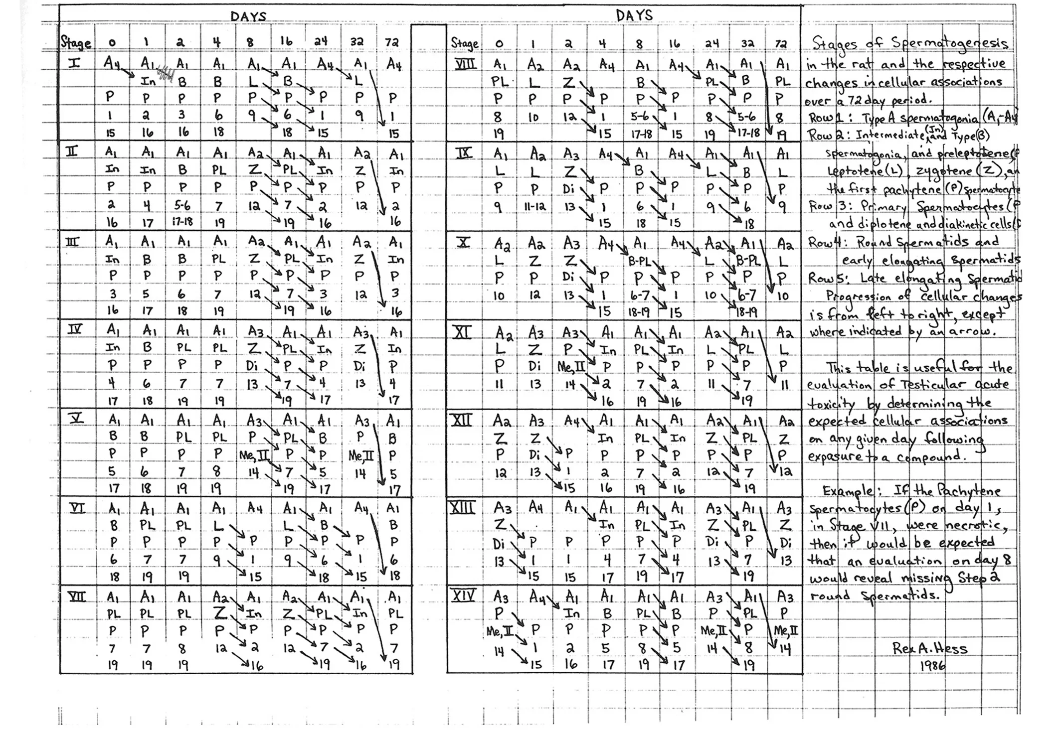
Before leaving the EPA for Illinois in 1986, I was involved in a toxicology study to determine the effects of 1,3-Dinitrobenzene on male reproduction. All animal exposures and tissue collections had been completed at the EPA, but histology needed work-up and my analysis of spermatogenesis was still in its infancy. After moving to Illinois, I was fortunate to continue the EPA collaboration and eventually completed analysis of the testis, but not without an intensive effort that required hand-written recordings of germ cell changes in each stage of rat spermatogenesis over time (Fig. 1). In 1988 we were able to use these kinetic data for the first time in a toxicology study, showing, for example, that “1,3-Dinitrobenzene does not appear to have a primary effect on the germ cells that are outside the blood-testis barrier (spermatogonia and preleptotene spermatocytes), because by day 16 these cell types would have become pachytene spermatocytes” (Hess et al. 1988).
Birth of a Computer Program
Step by step, research led me to the concept of a program called STAGES, which took shape between 1986 and 1987. The tedium of the manual work to track germ-cell progression through the stages in time was so cumbersome, inefficient, prone to error and difficult to teach that I dreamed of using the computer for this purpose. Therefore, I contacted the Head of the Department of Electrical and Computer Engineering at the University of Illinois, who introduced me to a PhD student in his department, Paul Chen. I designed the program on paper and, after showing him the collection of stages grid information and kinetics, Paul wrote the code in Turbo C for MS-DOS beginning in 1988. The program was distributed on floppy disks, extensively tested, and later described in a 1992 publication (Hess and Chen 1992). Today, Dr. Chen is Senior Director of Product Management at Komprise in San Francisco.
Windows and Mac Versions
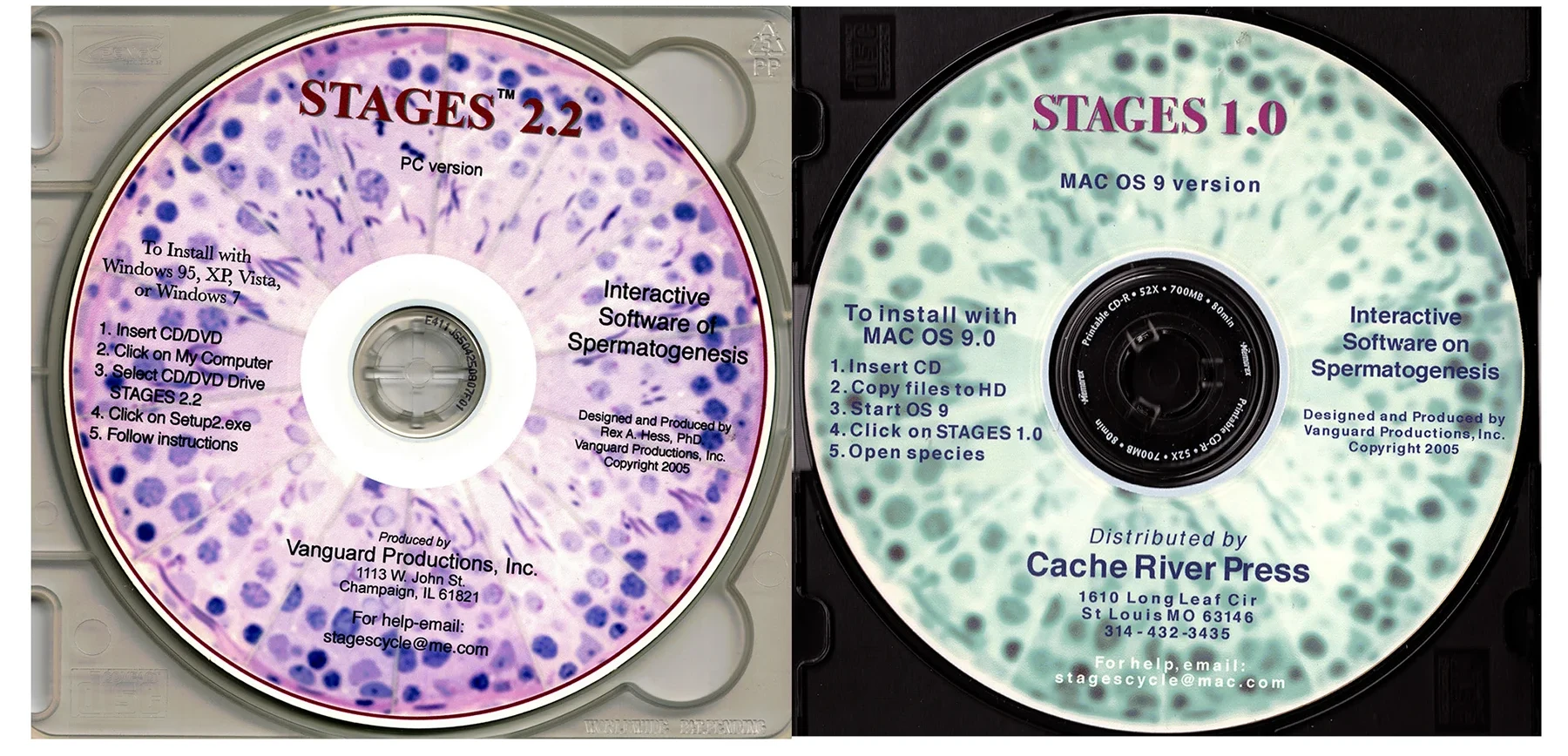
Soon, rapid changes in computing began to disrupt use of the program. Microsoft launched Windows 3.0 in 1990, making the DOS version harder to run. Therefore, between 1993 and 1994, I hired a veterinary student, David Scott, to rewrite STAGES in C++ for Windows, and in 1995 hired an undergraduate student, Daniel Pape, to code the Mac version. Figure 2 shows the 2005 versions. After acquiring full rights, title and ownership from the University of Illinois, Scott and Pape, I registered the program with the U.S. Copyright Office on March 17, 2006 (TX0006361724), with a 1995 publication date for the CD-ROM. Early in 1995, I sent a Windows version of STAGES to Dr. Yves Clermont of McGill University for review. I was honored to receive his hand-written letter of approval and later excellent suggestions, as he was co-author of the seminal 1952 paper that defined 14 stages in the rat seminiferous epithelial cycle using PAS staining of the acrosomic system (Leblond and Clermont 1952). In hindsight, his 1952 paper and personal encouragement were inspirational to the development of this computer program.
SpermatoStages™ Browser Version
The CD/DVD version, STAGES 2.2, was sold through Vanguard Productions, Inc. (Rex A. Hess, Proprietor) to scientists worldwide and later distributed also by Cache River Press (owned by my close friend and colleague, Dr. Lonnie Russell, before his passing in 2001). About 10 to 15 years ago, the program became obsolete, as it would not run on the new Operating Systems in both Windows and Mac computers. However, in 2026 I decided to continue developing the program, begun more than four decades earlier. STAGES was renamed SpermatoStages™ to distinguish it from other software with the same name. It was rewritten as a new program, with the prediction kernel ported line-by-line from C++ to JavaScript and designed to reproduce the original arithmetic. Its results match the Windows program (tested for several species). The web application, written in HTML, CSS, and JavaScript, runs entirely in the browser and was launched on July 22, 2026, and is now accessible at https://spermatostages.com. SpermatoStages™ is a web-based, interactive eBook and model of the cycle of the seminiferous epithelium. It provides a flexible stage-prediction algorithm, stage grid data for multiple species, breeds, and strains, adjustable kinetics, drawings, photographs, references, and notes.
Professor Emeritus
University of Illinois Urbana-Champaign
[email protected]
[email protected]
References
- Chapin RE, Morgan KT and Bus JS (1983). The morphogenesis of testicular degeneration induced in rats by orally administered 2,5-hexanedione. Exp Mol Pathol 38(2): 149–169. https://doi.org/10.1016/0014-4800(83)90082-5
- Chapin RE, Ross MD and Lamb JC (1984). Immersion Fixation Methods for Glycol Methacrylate-embedded Testes. Toxicologic Pathology 12(3): 221–227. https://doi.org/10.1177/019262338401200303
- Clermont Y, Leblond CP and Messier B (1959). Durée du cycle de l'épithélium séminal du rat. Arch Anat Microsc Morphol Exp 48(Suppl): 37–55.
- Hess RA (1990). Quantitative and qualitative characteristics of the stages and transitions in the cycle of the rat seminiferous epithelium: light microscopic observations of perfusion-fixed and plastic-embedded testes. Biol Reprod 43(3): 525–542. https://doi.org/10.1095/biolreprod43.3.525
- Hess RA and Chen P (1992). Computer tracking of germ cells in the cycle of the seminiferous epithelium and prediction of changes in cycle duration in animals commonly used in reproductive biology and toxicology. J Androl 13(3): 185–190. https://doi.org/10.1002/J.1939-4640.1992.TB00297.X
- Hess RA, Linder RE, Strader LF and Perreault SD (1988). Acute effects and long-term sequelae of 1,3-dinitrobenzene on male reproduction in the rat. II. Quantitative and qualitative histopathology of the testis. J Androl 9(5): 327–342. https://doi.org/10.1002/j.1939-4640.1988.tb01059.x
- Leblond CP and Clermont Y (1952). Definition of the stages of the cycle of the seminiferous epithelium in the rat. Ann NY Acad Sci 55: 548–573. https://doi.org/10.1111/j.1749-6632.1952.tb26576.x
- Oakberg EF (1956). Duration of spermatogenesis in the mouse and timing of stages of the cycle of the seminiferous epithelium. Am J Anat 99(3): 507–516. https://doi.org/10.1002/aja.1000990307
- Roosen-Runge EC (1952). Kinetics of spermatogenesis in mammals. Ann N Y Acad Sci 55(4): 574–584. https://doi.org/10.1111/j.1749-6632.1952.tb26577.x